Understanding the Anatomy of the Spine
Anatomy of the Spine (Lumbar, Cervical, Back, Vertebrae, Sacral) in Texas
Throughout my career, I have seen many patients with back pain, neck pain, pinched nerve, sciatica, arthritis of the back, etc that are left in the dark about the root cause of their problem.
Many have been reading through their X-ray or MRI reports full of medical jargon without help, often leading to frustration, fear, and confusion.
At NorTex, we emphasize patient education first and we want to be able to explain the source of the problem in layman’s terms.
The first step in reviewing any spine-related problems is to understand the anatomy of the spine and its structures.
We have put together a brief description of the spine as well as a video to help understand this rather complex and marvelous structure of the body. Sincerely, Oliver Ghalambor, MD
Overview
The spinal column is the body’s main support structure. Its thirty-three bones, called vertebrae, are divided into five regions: cervical, thoracic, lumbar, sacral, and coccygeal.
Cervical Region
The cervical region consists of seven vertebrae labeled C1 to C7. The first cervical vertebra is called the atlas. The second is called the axis. Together, the atlas and axis form the joint that connects the spine to the skull and allows the head to swivel and nod.
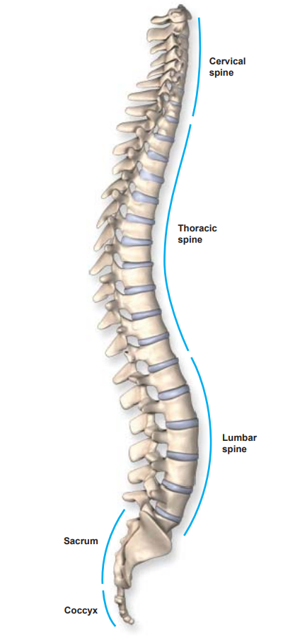
Thoracic Region
The thoracic region, located in the mid-back, consists of twelve vertebrae labeled T1 to T12. These vertebrae serve as attachment points for the ribcage.
Lumbar Region
The lumbar region, commonly called the lower back, consists of five vertebrae labeled L1 to L5. This is the main weight-bearing section of the spinal column.
Sacral region
The sacral region consists of five fused vertebrae labeled S1 to S5. These vertebrae form a solid mass of bone, called the sacrum, which provides the attachment point for the pelvis.
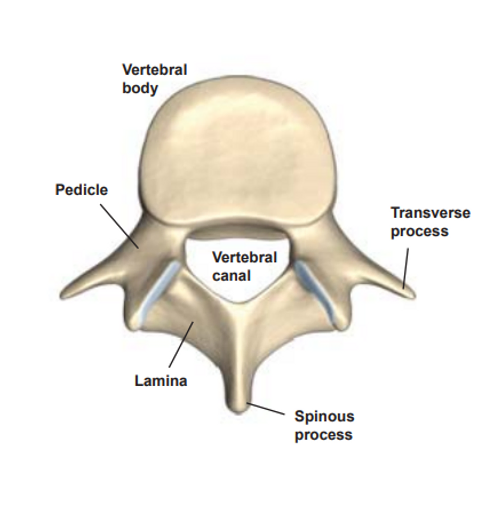
Vertebral Canal
Behind the vertebral body is the vertebral canal. The spinal cord travels through this channel.
NorTex Spine & Joint Institute
Is proud to be the leader of non-surgical treatments for a variety of spine-related problems offered by Dr. Ghalambor, Harvard Trained, Fellowship Trained, and Board Certified Specialist.
We offer consultations and treatments in our affiliated clinics in Plano, McKinney, Frisco, Lewisville, Wylie, Celina, Garland, Allen, Addison, and Dallas in Texas.
Want to talk more about your Back Pain, Neck Pain, or other Spine related problems?
Call us today at 972-872-8408
Coccygeal Region
The coccygeal region, commonly called the tailbone, consists of four small vertebrae. These tiny bones may be fused or separate. Together they form the coccyx, an attachment point for various muscles, tendons and ligaments. The coccyx also helps support the body when a person is sitting.
Vertebrae
Altogether, the vertebrae of the spine’s five regions support the weight of the body and protect the spinal cord and nerve roots. Each individual vertebra has a complex set of structures necessary to the overall function of the spine.
Watch Dr. Ghalambor and Bob discuss Anatomy
Dr. Ghalambor discussing Anatomy Of the Spine
Vertebral Body
The main structure of a vertebra is the vertebral body — a cylinder-shaped section of bone at the front of the vertebra. It is the main weight-bearing section of the vertebra.
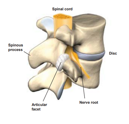
Spinal Cord
The spinal cord is the main bundle of nerve fibers connecting the brain to the rest of the body. The spinal cord ends near the L1 and L2 vertebrae, where it divides into bundles of nerve roots called the cauda equina.
Nerve Roots
Exiting the sides of the spine are nerve roots, thick nerve branches that transmit signals between the spinal cord and the other parts of the body.
Pedicles
On either side of the vertebral canal are pedicle bones, which connect the vertebral body to the lamina.
Dr. Ghalambor's Overview Of the Lumbar Spine
Dr. Ghalambor's Overview Of the Cervical Spine
Lamina
The lamina create the outer wall of the vertebral canal, covering and protecting the spinal cord.
Spinous Process
Protruding from the back of the lamina is the spinous process. It provides an attachment point for muscles and ligaments that move and stabilize the vertebrae.
Transverse Processes
Transverse processes protrude from the sides of each vertebra. Muscles and ligaments that move and stabilize the vertebrae attach to the transverse processes.
Articular Facet
The articular facets form the joints where each vertebra connects with the vertebrae above and below it. Each vertebra has four facets (two superior facets and two inferior facets). The facet joints have a covering of cartilage, which allows movement.
Intervertebral Disc
Between the vertebral bodies are the tough, elastic spinal discs. They provide a flexible cushion, allowing the vertebrae to bend and twist. Each disc has a tough outer wall called the annulus fibrosus and a soft interior called the nucleus pulposus.