
Platelet-rich plasma (PRP) promotes tissue healing by delivering a concentrated, autologous pulse of platelets and plasma-borne factors that alter the local microenvironment to favor repair — accelerating hemostasis, modulating inflammation, driving angiogenesis, and stimulating cell proliferation and matrix synthesis. According to a comprehensive PMC mechanistic review, these actions work in concert to overcome the sluggish intrinsic repair response common in chronic and degenerative tissues. The primary mechanisms include:
- Hemostasis and cell recruitment: Activated platelets form a fibrin scaffold and release chemotactic signals that draw mesenchymal stem cells (MSCs) and fibroblasts to the injury site.
- Growth factor release: Alpha-granule contents — PDGF, TGF-β, VEGF, and EGF — are secreted rapidly, initiating and sustaining the proliferative repair cascade.
- Immunomodulation: PRP shifts the cytokine environment away from pro-inflammatory mediators (IL-6, TNF-α) toward an anti-inflammatory, anabolic state.
- Angiogenesis: VEGF and PDGF together drive new capillary formation, restoring oxygen and nutrient delivery to damaged tissue.
- Matrix remodeling: Fibroblast activation and collagen synthesis rebuild the extracellular matrix, restoring structural integrity.
- Analgesia: Platelet-derived serotonin and improved tissue oxygenation reduce nociceptive signaling, contributing to pain relief.
Table of Contents
- What is PRP and where is it used clinically?
- How PRP is prepared and why the variations matter
- What PRP actually does inside injured tissue
- What the clinical evidence shows by indication
- What happens during a PRP procedure and when to expect results
- Safety, side effects, and contraindications
- Why PRP outcomes vary between patients and trials
- Limitations and open questions in the current evidence
- How Nortexspineandjoint approaches PRP in clinical practice
- Key Takeaways
- What I tell patients about PRP — a clinical perspective
- Nortexspineandjoint’s PRP services and how to take the next step
- FAQ
- Primary sources and further reading
What is PRP and where is it used clinically?
Platelet-rich plasma is an autologous blood concentrate prepared at the point of care. A small volume of your own blood is processed to yield a fraction with a platelet concentration substantially above baseline whole blood, along with plasma proteins that contribute independently to cellular signaling. The result is a biologic preparation that carries none of the immunogenic risk associated with donor-derived products.
Clinicians classify PRP along several dimensions: leukocyte-rich (LR-PRP) versus leukocyte-poor (LP-PRP), platelet concentration, and fibrin architecture. Each classification reflects a different biological profile and, consequently, a different clinical application. Protein-rich PRP matrices (PR-PRP) represent a further refinement, combining concentrated platelets with a small volume of platelet-poor plasma (PPP) to create a sustained-release fibrin scaffold.
The clinical applications span multiple specialties. In orthopedics, PRP is most commonly used for tendinopathy, knee and hip osteoarthritis, and ligament or tendon injuries. Dermatologists use it for androgenetic alopecia and skin rejuvenation. Wound care specialists apply it to chronic diabetic wounds and surgical incisions. Dental and maxillofacial surgeons use platelet-rich fibrin membranes to support periodontal regeneration and bone defect repair. For patients considering PRP for joint conditions, Nortexspineandjoint’s clinical guide to PRP for knees provides a practical overview of the evidence supporting its use.
How PRP is prepared and why the variations matter
Preparation follows a consistent sequence, though the details at each step produce meaningfully different products:
- Blood draw: Typically 10–60 mL of venous blood, collected into anticoagulant tubes (sodium citrate or ACD-A) to prevent premature clotting.
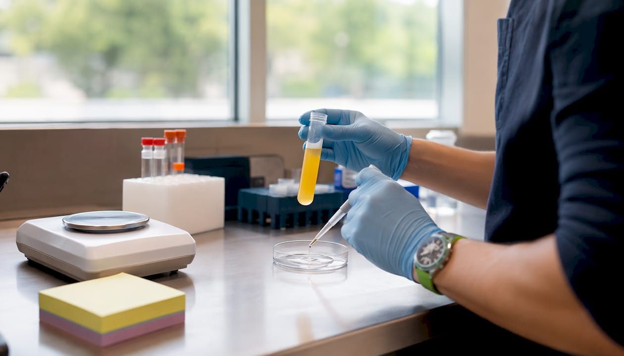
- Centrifugation: A single-spin protocol separates red cells from the buffy coat and plasma; a double-spin or buffy-coat method further concentrates platelets. The spin speed and duration determine the final platelet yield and leukocyte content.
- Fraction selection: The clinician draws off the platelet-rich layer. In LP-PRP protocols, the buffy coat (leukocyte layer) is excluded; in LR-PRP, it is included.
- Optional activation: Calcium chloride or thrombin can be added to trigger degranulation before injection, creating a gel or matrix rather than a liquid suspension.
- Delivery: Injected directly into the target tissue, often under ultrasound guidance for intra-articular or tendon applications.
LR-PRP vs. LP-PRP is not a trivial distinction. Leukocytes carry their own cytokine payload, which can amplify the initial inflammatory signal — potentially beneficial in tendon repair but possibly counterproductive in joint environments where cartilage is sensitive to prolonged inflammation. LP-PRP is generally preferred for intra-articular injections; LR-PRP is more commonly used for tendinopathies and wound healing.
Platelet concentration matters in a dose-dependent way. Many practitioners reference a threshold near ~1.5 million platelets/µL as a practical target for effective tissue remodeling, though excessively high concentrations may paradoxically inhibit angiogenesis. Plasma proteins also contribute: IGF-1 and other factors present in PPP support cellular proliferation and anti-fibrotic signaling in ways that platelet count alone does not capture, which is the rationale behind PR-PRP matrix strategies.
Pro Tip: No single PRP device or protocol is universally standardized. The same brand name can produce substantially different platelet concentrations depending on the operator’s technique, the patient’s baseline platelet count, and the centrifuge settings used. When reviewing clinical trial results, always check whether the study reports platelet concentration — and be cautious about generalizing findings from one protocol to another.
What PRP actually does inside injured tissue
Understanding the cellular sequence helps clarify why PRP works in some tissues and why results vary. The process unfolds in overlapping phases.
Immediate actions (minutes to hours). When PRP contacts tissue, platelets activate and degranulate. Alpha granules release PDGF, TGF-β, VEGF, and EGF within minutes; more than 95% of pre-synthesized growth factors are secreted within the first hour. Dense granules release serotonin, which contributes to local analgesia. The fibrin matrix formed during activation serves as a physical scaffold, trapping platelets and allowing a slower, sustained release of mediators over the following days.
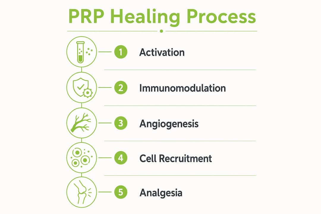
Immunomodulation (hours to days). PRP reduces pro-inflammatory cytokines such as IL-6 and TNF-α while increasing anti-inflammatory proteins in the joint or tissue environment. This shift drives macrophages from a pro-inflammatory M1 phenotype toward a reparative M2 phenotype, resolving the chronic inflammatory state that characterizes degenerative conditions. In tissues where the normal repair cycle has stalled, this cytokine reset is often the most clinically meaningful early event.
Angiogenesis (days to weeks). VEGF drives endothelial cell proliferation and capillary sprouting; PDGF stabilizes new vessel walls by recruiting pericytes. Restoring microvascularity is particularly critical in tendons and cartilage-adjacent tissues, which are inherently hypovascular. Platelet dosing and bioformulation strategies strongly influence angiogenic responses, which is one reason why underdosed preparations often fail to produce durable clinical benefit.
Cell recruitment and matrix synthesis (days to weeks). PDGF acts as a chemotactic signal for MSCs, fibroblasts, and osteoblasts. Once recruited, these cells proliferate and begin synthesizing type I collagen and other extracellular matrix components. Exosome-mediated signaling from activated platelets adds another layer of paracrine communication that amplifies the regenerative response. PR-PRP matrices, which retain proteins and cells in a fibrin scaffold that undergoes gradual fibrinolysis, extend this window of mediator availability beyond what a liquid PRP injection provides.
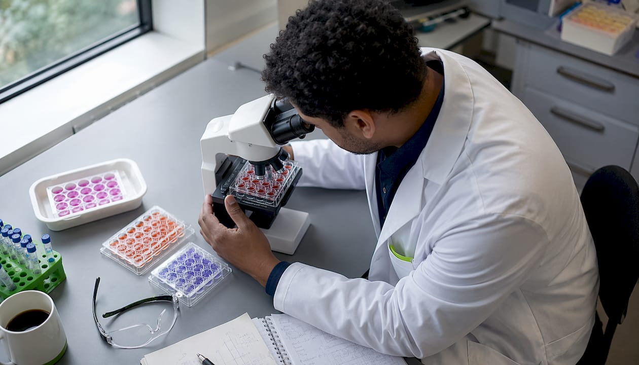
Analgesia. Pain relief from PRP is partly direct (platelet-derived serotonin modulates nociceptor activity) and partly indirect: as inflammation resolves and tissue oxygenation improves through neo-angiogenesis, the nociceptive load decreases. Clinical observations and some randomized controlled trials report reduced pain and improved function following PRP in both surgical and non-surgical contexts.
| Growth Factor / Mediator | Primary Action | Repair Outcome |
|---|---|---|
| PDGF (Platelet-Derived Growth Factor) | Chemotaxis of MSCs, fibroblasts, osteoblasts; mitogen for mesenchymal cells | Cell recruitment, angiogenesis, fibrous tissue formation |
| TGF-β (Transforming Growth Factor-β) | Stimulates collagen synthesis; promotes angiogenesis and ECM production | Matrix remodeling, scar regulation, tissue strengthening |
| VEGF (Vascular Endothelial Growth Factor) | Endothelial cell proliferation and migration | Capillary formation, improved tissue oxygenation |
| EGF (Epidermal Growth Factor) | Mitogen for fibroblasts, keratinocytes, endothelial cells | Re-epithelialization, wound closure |
| IGF-1 (Insulin-like Growth Factor-1) | Induces proliferation, differentiation, and hypertrophy of multiple cell lines | Cellular proliferation, anti-fibrotic signaling |
| Serotonin (5-HT, dense granules) | Modulates nociceptor activity | Direct analgesia, vasoregulation |
What the clinical evidence shows by indication
The strength of evidence varies considerably across indications. The table below summarizes the current picture.
| Indication | Typical Benefit | Time to Improvement | Evidence Quality |
|---|---|---|---|
| Knee osteoarthritis | Clinically meaningful pain and function improvement; high-platelet PRP superior to low-platelet PRP at 12 months | 6–12 weeks; sustained to 6–12 months | Moderate to high (multiple RCTs, meta-analyses) |
| Skin and wound healing | Improved wound closure, granulation tissue, reduced inflammation | Days to weeks | Moderate (preclinical and clinical studies) |
| Tendinopathy | Modest to moderate benefit in some trials; results inconsistent across prep methods | 6–12 weeks | Low to moderate (heterogeneous trials) |
| Hair restoration (androgenetic alopecia) | Increased hair density in several RCTs; some trials show limited effect | 3–6 months | Low to moderate |
| Post-surgical adjunct | Reduced postoperative pain in some contexts; perioperative use supported | Immediate to weeks | Moderate (indication-specific) |
Knee osteoarthritis has the most consistent evidence base. Meta-analyses show that trials using ≥40 mL blood draw volumes — a practical proxy for higher platelet yield — report significantly improved WOMAC and VAS outcomes compared with lower-volume trials. This dose-response signal is one of the more actionable findings for clinicians selecting protocols.
Wound healing evidence is supported by both preclinical and clinical data showing improved re-epithelialization, granulation tissue formation, and reduced inflammation with PRP. Tendinopathy and hair restoration trials are more mixed, largely because preparation heterogeneity makes cross-study comparison unreliable.
What happens during a PRP procedure and when to expect results
Understanding the appointment sequence helps set realistic expectations. Here is the typical clinical workflow:
- Consultation and candidacy review. Your clinician reviews imaging, medical history, and treatment goals to confirm PRP is appropriate for your condition.
- Blood draw. A venous blood sample is collected, typically ranging from 15–60 mL depending on the protocol and target platelet dose.
- PRP preparation. The blood is centrifuged at the point of care. Preparation takes roughly 15–20 minutes.
- Injection. PRP is delivered to the target tissue. For intra-articular injections (knee, hip) and tendon applications, ultrasound guidance improves accuracy and safety.
- Immediate aftercare. Avoid anti-inflammatory medications (NSAIDs and corticosteroids) for at least one to two weeks post-injection, as they can blunt the intended inflammatory-repair signal. Light activity is generally encouraged; high-impact loading is restricted initially.
- Follow-up. Most clinicians schedule a reassessment at 6–12 weeks to evaluate early response, with a more definitive outcome assessment at 3–6 months.
Timeline for biological and clinical response:
- Hours to days: Hemostasis, fibrin scaffold formation, early growth factor release, initial inflammatory modulation.
- Days to weeks: Immunomodulation, macrophage phenotype shift, angiogenesis begins.
- Weeks to months: Active matrix remodeling, collagen synthesis, measurable functional improvement.
- 3–6 months: Primary window for clinical outcome assessment in most orthopedic indications.
- 6–12 months: Sustained benefit assessment; high-platelet knee OA protocols show durable improvement at this horizon.
For a detailed look at what to expect at your first visit, Nortexspineandjoint’s first-visit guide covers the consultation process and realistic timelines in practical terms.
Pro Tip: PRP works best as part of a structured rehabilitation program, not as a standalone injection. The growth factors released by PRP create a biological window of opportunity for tissue remodeling — but that window needs to be paired with progressive loading and physical therapy to translate cellular repair into functional recovery. Patients who combine PRP with a supervised rehab protocol consistently report better outcomes than those who rest passively.
Safety, side effects, and contraindications
Because PRP is autologous, the immunogenic risk is minimal. The most common adverse events are local and transient:
- Injection-site pain and soreness, typically peaking at 24–72 hours post-procedure
- Transient swelling or stiffness at the injection site
- Small hematoma at the blood draw or injection site
- Brief exacerbation of symptoms before improvement, particularly in the first one to two weeks
Serious complications are rare. Infection is possible with any injection procedure but is uncommon when standard sterile technique is followed. Some patients experience no durable benefit, which is a clinical outcome rather than a safety event.
Contraindications to discuss with your clinician include:
- Active infection at or near the injection site
- Platelet function disorders or severe thrombocytopenia
- Uncontrolled systemic illness (active malignancy, sepsis)
- Anticoagulation therapy (warfarin, direct oral anticoagulants) — management requires individual assessment
- Pregnancy, where applicable depending on the indication
The pros and cons of PRP injections are worth reviewing carefully with your treating clinician, who can weigh your specific medical history, current medications, and treatment goals before proceeding.
This article provides general educational information, not medical advice. Confirm the appropriateness of PRP for your specific situation with a qualified clinician.
Why PRP outcomes vary between patients and trials
Results differ because multiple variables interact to determine the final biological response. These fall into three categories:
Patient factors:
- Age and baseline tissue vascularity (older patients and those with diabetes or peripheral vascular disease tend to have impaired angiogenic responses)
- Smoking, which reduces platelet function and tissue oxygenation
- Severity of the underlying condition (early-to-moderate OA responds better than end-stage disease)
- Baseline platelet count, which directly affects the yield achievable from a given blood draw volume
Product factors:
- Total platelet dose and concentration (dose-dependent effects are well-documented)
- Leukocyte content (LR vs. LP formulations produce different cytokine environments)
- Inclusion of PPP and plasma proteins such as IGF-1, which contribute to proliferative signaling
- Activation method (exogenous thrombin or calcium vs. endogenous activation at the tissue site)
Technique factors:
- Injection accuracy, particularly for intra-articular and tendon targets
- Use of ultrasound guidance, which reduces extra-articular misplacement
- Volume delivered and number of injections in a series
Laboratory and reporting variability compounds these differences across studies. When platelet-dose data are missing from a trial, blood draw volume of ≥40 mL can serve as a practical proxy for achieving higher platelet yields, and trials using this threshold have shown consistently better outcomes in knee OA. Readers interested in how peptide-based adjuncts interact with these repair pathways may find the role of peptides in muscle repair a useful parallel reference.
Limitations and open questions in the current evidence
Transparency about evidence gaps is part of responsible clinical communication. The main limitations are:
- Preparation heterogeneity: No universal PRP standard exists. Different devices, spin protocols, and leukocyte strategies produce products with substantially different biological profiles, making cross-trial comparisons unreliable.
- Small and underpowered trials: Many early PRP studies enrolled fewer than 50 patients per arm, limiting statistical power and generalizability.
- Short follow-up: A significant proportion of trials report outcomes only to 3–6 months, leaving long-term disease-modification potential largely unanswered.
- Variable outcome measures: Trials use different pain scales, functional scores, and imaging endpoints, further complicating meta-analytic synthesis.
Open scientific questions include the optimal platelet dose for each indication, the precise role of leukocytes in different tissue environments, the best activation strategy, and whether PRP combined with cell-based therapies or scaffolds produces additive benefit. The MIBO (Minimum Information for studies evaluating Biologics in Orthopaedics) reporting guidelines represent an effort to standardize what trials must disclose, but adoption remains inconsistent.
Shared decision-making with a clinician who can interpret the evidence in the context of your specific diagnosis, imaging findings, and treatment history is the most reliable path to a well-calibrated decision.
How Nortexspineandjoint approaches PRP in clinical practice
Many patients who arrive at Nortexspineandjoint have already tried physical therapy, oral anti-inflammatories, and one or more corticosteroid injections without lasting relief. That pattern — partial responders to conservative care with early-to-moderate structural changes on imaging — represents the most common PRP candidate seen in practice.
Before recommending PRP, the clinical team reviews recent imaging (X-ray and MRI where indicated), confirms that the structural findings align with the patient’s reported symptoms, and discusses realistic outcome expectations. Candidacy is not determined by diagnosis alone. A patient with moderate knee OA and intact joint space is a different candidate than one with bone-on-bone changes, and the conversation reflects that distinction.
Blood draw volumes are selected with platelet yield in mind. For most orthopedic indications, protocols targeting the ≥40 mL threshold are preferred, consistent with the meta-analytic evidence linking higher draw volumes to better outcomes. Ultrasound guidance is used routinely for intra-articular and tendon injections to confirm accurate placement. Platelet counts are documented when available to support outcome tracking.
Patients typically see early signs of response — reduced soreness, improved range of motion — within the first four to six weeks. More meaningful functional gains generally emerge at the three-month mark. For knee and hip OA, the six-month assessment is where the clinical picture becomes clearest.
Questions to ask your clinician before PRP treatment:
- What platelet concentration or blood draw volume will you use, and why?
- Will the injection be guided by ultrasound?
- What rehabilitation protocol do you recommend alongside the injection?
- How will we define a successful outcome, and at what timepoint?
- What are the next steps if PRP does not produce adequate improvement?
Nortexspineandjoint serves patients across North Dallas, including Allen, Garland, McKinney, and Plano, Texas. The practice’s approach to PRP is grounded in the same evidence reviewed in this article, applied to individual patient presentations rather than a one-size protocol.
Key Takeaways
PRP promotes healing through a dose-dependent, multi-mechanism biological response that is strongest when platelet concentration, leukocyte content, and injection technique are matched to the target tissue and indication.
| Point | Details |
|---|---|
| Core mechanism | PRP delivers concentrated growth factors (PDGF, TGF-β, VEGF, EGF) that trigger cell recruitment, angiogenesis, and matrix remodeling in injured tissue. |
| Dose matters | High-platelet PRP outperforms low-platelet PRP in knee OA trials; larger blood draw volumes correlate with better patient-reported outcomes. |
| Strongest evidence | Knee osteoarthritis has the most consistent evidence base, with clinically meaningful improvement sustained to 12 months in high-platelet trials. |
| Safety profile | PRP is autologous and generally well-tolerated; common adverse events are local and transient, with serious complications rare. |
| Nortexspineandjoint | Nortexspineandjoint evaluates PRP candidates based on imaging alignment, treatment history, and realistic outcome goals, using ultrasound guidance and evidence-based dosing protocols. |
What I tell patients about PRP — a clinical perspective
Patients often ask whether PRP is “worth it.” My honest answer is that it depends on what you are treating, how the product is prepared, and what you do afterward.
For knee osteoarthritis in the early-to-moderate range, the evidence is genuinely encouraging. Multiple meta-analyses now show that high-platelet preparations produce clinically meaningful improvements in pain and function that persist to 12 months. That is a meaningful result for a patient who has exhausted conservative options and wants to avoid or delay surgery. I tell those patients that PRP is a reasonable next step, with realistic expectations: most will notice gradual improvement over six to twelve weeks, not overnight relief.
For tendinopathies, the picture is more nuanced. Some patients respond well; others do not. The variability is real, and I think it reflects preparation differences as much as biology. When a patient asks whether PRP will definitely help their Achilles tendinopathy, I explain that the evidence supports a trial, particularly when combined with eccentric loading rehabilitation, but I cannot guarantee a specific outcome.
The question I hear most often is whether the injection hurts. It is uncomfortable, not unbearable. The post-injection soreness in the first 48–72 hours is typically the most significant discomfort, and it is a sign that the biological process has started. Patients who expect that soreness and plan for it tolerate the procedure well.
PRP fits into a broader care plan, not a replacement for one. When structural damage is severe or when a patient’s goals require a level of function that biologic therapy alone cannot restore, surgical referral is still appropriate. PRP does not change that calculus. What it can do, in the right patient with the right preparation, is meaningfully extend the window of non-surgical management.
Nortexspineandjoint’s PRP services and how to take the next step
Patients who have read this far are usually past the “is PRP real?” question and into the more practical one: “Is it right for me?” That is exactly the conversation a consultation at Nortexspineandjoint is designed to have.
The clinic’s PRP therapy service covers the full evaluation: a review of your imaging and treatment history, a candidacy assessment based on your specific diagnosis and goals, a discussion of the preparation protocol and expected timeline, and a clear explanation of what the evidence supports for your indication. There are no blanket promises. What you will leave with is a realistic picture of whether PRP fits your situation and what a structured treatment plan would look like.
For patients with knee or hip osteoarthritis specifically, Nortexspineandjoint’s regenerative medicine program integrates PRP with rehabilitation planning and, where appropriate, other biologic options. To schedule a consultation at the Allen, Garland, McKinney, or Plano, Texas locations, contact the clinic directly through the website.
FAQ
What does PRP do biologically to promote healing?
PRP activates platelets that release growth factors (PDGF, TGF-β, VEGF, EGF) from alpha granules, triggering cell recruitment, angiogenesis, and matrix remodeling while modulating inflammation through cytokine shifts that favor tissue repair over chronic inflammation.
How many PRP injections are typically needed?
Most orthopedic protocols involve one to three injections, spaced several weeks apart, though the number depends on the indication, the patient’s response, and the clinician’s protocol. A single well-dosed injection is standard for many knee OA protocols.
When will I feel improvement after a PRP injection?
Early changes in soreness and range of motion often appear within four to six weeks; more meaningful functional gains typically emerge at the three-month mark, with the clearest clinical picture at six months for most orthopedic indications.
Is PRP safe, and are there people who should not receive it?
PRP is autologous and generally well-tolerated, with common side effects limited to local, transient soreness and swelling. Contraindications include active infection at the injection site, platelet function disorders, severe thrombocytopenia, and uncontrolled systemic illness.
Does platelet concentration affect how well PRP works?
Yes. High-platelet PRP formulations produce superior pain relief and longer-lasting functional improvement in knee OA compared with low-platelet preparations, and blood draw volumes of ≥40 mL correlate with better patient-reported outcomes across trials.
Primary sources and further reading
- Platelet-Rich Plasma (PRP): Molecular Mechanisms, Actions and Clinical Applications — PMC — Comprehensive mechanistic and clinical review; primary source for growth factor biology and analgesia mechanisms.
- PRP Injections for Knee Osteoarthritis: Platelet Concentration and Clinical Improvement — PMC — Meta-analysis comparing high- vs. low-platelet PRP in knee OA.
- Autologous PRP vs. Hyaluronic Acid, Corticosteroids, or Saline for Knee OA: Blood Draw Volume as Proxy — International Orthopaedics — Systematic review supporting ≥40 mL blood draw as a practical dosing proxy.
- Profound Properties of Protein-Rich, Platelet-Rich Plasma Matrices — MDPI — Covers PR-PRP matrix biology and sustained-release mechanisms.
- Angiogenesis and Tissue Repair Depend on Platelet Dosing and Bioformulation — Biomedicines (MDPI) — Narrative review on angiogenic mechanisms and dosing strategy.
- Platelet-Therapeutics to Improve Tissue Regeneration and Wound Healing — PMC — Broad review of platelet physiology, preparation methods, and clinical applications.
- Platelet-Rich Plasma Peptides: Key for Regeneration — PMC — Background on platelet-derived growth factor biology and dose-response relationships.
- Frontiers in Cell and Developmental Biology: PRP Mechanism Review — Contemporary framing of PRP as a concentrated repair signal for degenerative tissues.
- ClinicalTrials.gov — Search for active PRP trials by indication (e.g., “platelet-rich plasma knee osteoarthritis”) to find ongoing studies relevant to your condition.
Recommended
- What Makes PRP Therapy So Effective? – Nortex | Pain Management
- Why Consider PRP for Knees: A Clinical Guide
- How PRP Injections Can Speed Up Your Injury Recovery – Nortex | Pain Management | Allen, Garland, McKinney & Plano Texas
- PRP Therapy: A Breakthrough in Treating Chronic Wounds – Nortex | Pain Management | Allen, Garland, McKinney & Plano Texas