
Joint repair refers to the set of surgical and biologic procedures designed to restore or preserve a damaged joint surface rather than replace the joint entirely. If you are trying to understand which approach fits your situation, here is the short version:
- Non-surgical biologics (PRP, BMAC, MFAT/AMAT): best for early-to-moderate cartilage symptoms, focal tendinopathy, or early osteoarthritis where structural damage is limited
- Marrow stimulation / microfracture: a surgical option for small focal cartilage defects, typically in younger, lower-demand patients
- Cell-based cartilage restoration (ACI/MACI): staged procedures for medium-to-large contained defects in patients willing to commit to a long rehabilitation
- Osteochondral grafting (autograft/OAT, allograft): structural bone-and-cartilage transfer for defects where immediate hyaline cartilage is needed
- Osteotomy: alignment correction to protect a repair or delay arthroplasty
- Total joint replacement: the appropriate endpoint when degeneration is widespread and repair is no longer viable
The right choice depends on lesion size, joint alignment, your age and activity goals, prior surgeries, and how much rehabilitation time you can commit to. Patients with specific, positive expectations about outcomes are nearly twice as likely to respond well at 6–12 months, which means preparation and honest goal-setting matter as much as the procedure itself.
Table of Contents
- What joint repair actually means — and how it differs from replacement
- Non-surgical biologic options: what PRP, BMAC, and MFAT can and cannot do
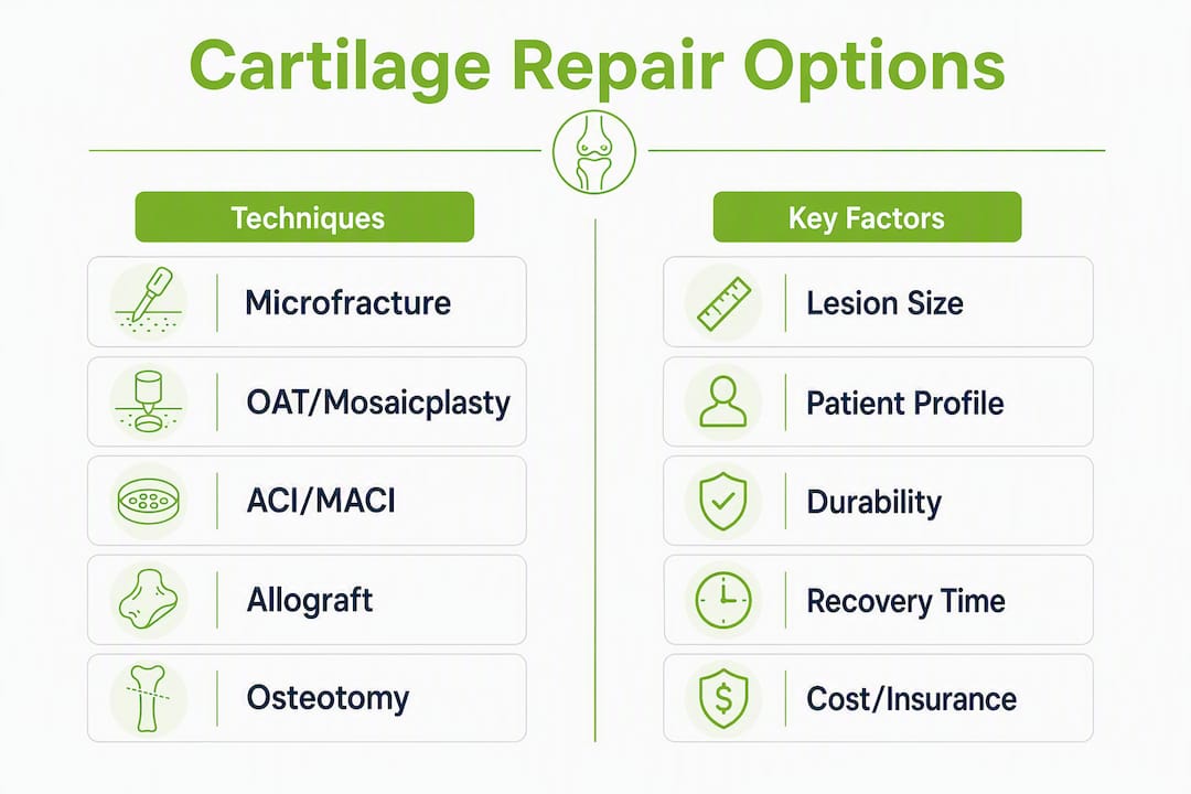
- Surgical cartilage-repair techniques: how the main options compare
- What emerging research is actually showing
- How clinicians decide which repair option fits you
- What to realistically expect: outcomes, durability, and rehab timelines
- Risks, complications, and why repairs sometimes fail
- Costs and insurance: what is typically covered in the U.S.
- When to see a specialist and how to prepare for that first visit
- Key Takeaways
- A clinician’s perspective on choosing the right path
- Nortexspineandjoint’s approach to joint repair evaluation
- Useful sources for further reading
- FAQ
What joint repair actually means — and how it differs from replacement
The term “joint repair” is used loosely, and that creates real confusion. Clinically, it describes procedures that preserve your native joint by restoring the cartilage surface, correcting alignment, or reinforcing the joint’s structural integrity. Joint replacement (arthroplasty) removes the damaged surfaces entirely and substitutes metal and plastic components. These are fundamentally different goals with different candidacy criteria, recovery demands, and long-term trajectories.
Cartilage restoration sits in the middle of that spectrum. The goal is to rebuild or regenerate the articular surface so the joint can function with reduced pain and improved mechanics, ideally delaying or avoiding arthroplasty. This is most appropriate for focal cartilage defects in younger or more active patients whose surrounding cartilage and bone are still in reasonable condition. Widespread, end-stage osteoarthritis with global joint degeneration is generally not a repair scenario.
One common mislabel worth correcting: arthroscopic debridement and lavage are sometimes called “repair,” but they do not restore cartilage. They remove loose tissue and may provide temporary symptom relief, but they do not address the underlying structural deficit. Knowing this distinction matters when you are evaluating what a proposed procedure will actually accomplish.
Non-surgical strategies and joint preservation techniques are also part of this continuum. Evidence shows that first-line conservative care can reduce willingness to undergo joint surgery by 30–45% at three months, and this effect is partially sustained at twelve months. That is not a reason to avoid surgery when it is indicated. It is a reason to take conservative care seriously before escalating.
Non-surgical biologic options: what PRP, BMAC, and MFAT can and cannot do
Orthobiologics are substances derived from your own body that are concentrated and injected into a damaged joint to modulate inflammation, support tissue healing, or provide a cellular environment for repair. They are not a replacement for structural procedures when significant cartilage loss is present, but they occupy an important role in the treatment spectrum.
Platelet-rich plasma (PRP) is prepared by drawing a small volume of your blood, centrifuging it to concentrate the platelets, and injecting the resulting plasma into the affected joint or tendon. Platelets release growth factors that can reduce inflammation and support soft-tissue healing. PRP is most commonly used for tendinopathy, early-to-moderate knee osteoarthritis, and as an adjunct to surgical procedures. The evidence supports short-term symptomatic improvement in many patients, but protocols vary widely across clinics, and structural imaging outcomes remain inconsistent. Leukocyte content, platelet concentration, and injection frequency all affect results, which is why standardization matters.
BMAC (bone-marrow aspirate concentrate) is drawn from the iliac crest under local anesthesia, concentrated at the point of care, and injected into the target joint. It contains mesenchymal stem cells, growth factors, and anti-inflammatory cytokines. BMAC differs from PRP in that it introduces a broader cellular population, including cells with potential to support cartilage repair. Candidate scenarios typically involve more significant cartilage damage than PRP alone would address, or patients who want a more potent biologic option before committing to surgery. You can learn more about bone marrow stem cell options and how they are evaluated at the clinic level.
MFAT / AMAT (microfragmented adipose tissue) is harvested from a small fat sample, typically from the abdomen or flank, processed to preserve the stromal vascular fraction, and injected into the joint. The proposed advantage is that adipose tissue contains a high concentration of regenerative cells within an intact extracellular matrix scaffold. Current evidence shows symptomatic benefit in early studies, but long-term structural durability data are still limited, and this approach remains investigational for most indications.
A note on “stem cell therapy” as marketed: In clinical practice, most point-of-care biologic procedures (BMAC, MFAT) contain a small fraction of mesenchymal stem cells alongside many other cell types. True autologous cell therapies involving expanded or engineered cell populations are a separate category, generally available only through FDA-regulated clinical trials. When a clinic markets “stem cell injections,” ask specifically what the preparation contains and whether it is a point-of-care concentrate or an investigational cell construct.
- PRP: best for tendinopathy, early OA, post-procedure adjunct
- BMAC: broader cellular profile, suited to more significant cartilage involvement
- MFAT/AMAT: promising early data, still investigational for structural repair
- All three: short-term symptom relief is common; structural durability evidence remains inconsistent
Pro Tip: When evaluating a clinic’s biologic program, ask for documented cell counts or platelet concentration data from their preparation process, a defined injection protocol (number of injections, interval), imaging follow-up at 3–6 months, and a coordinated rehabilitation plan. Clinics that cannot provide these specifics are operating without the documentation needed to interpret your outcome.
Surgical cartilage-repair techniques: how the main options compare
When biologics are insufficient or the defect is too large for injection-based management, surgical repair becomes the appropriate next step. Each technique has a specific lesion profile it is designed for, and applying a technique outside its intended constraints is the most common cause of premature failure.
Microfracture involves creating small perforations in the subchondral bone beneath a cartilage defect to allow marrow elements, including stem cells and growth factors, to fill the defect and form a fibrocartilage repair tissue. It is most appropriate for small defects (generally under 2 cm²) in younger patients with good bone quality. Recovery typically involves 6–8 weeks of protected weight-bearing, with return to sport at 4–6 months. The limitation is that fibrocartilage is mechanically inferior to native hyaline cartilage, and durability beyond 5 years is variable, particularly in higher-demand patients.
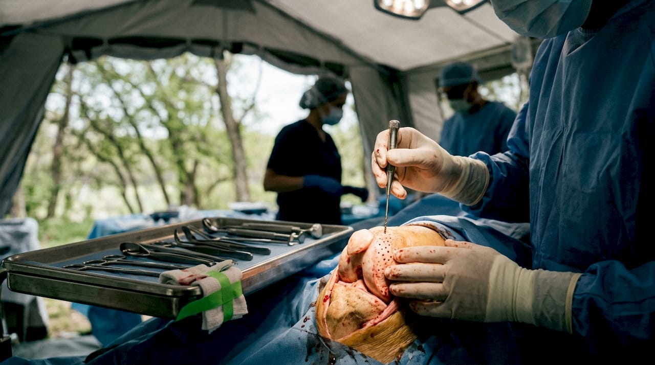
OAT / mosaicplasty (osteochondral autograft transfer) harvests cylindrical plugs of bone and cartilage from a low-load area of the same joint and transfers them to fill the defect. The advantage is immediate structural hyaline cartilage restoration in a single-stage procedure. It is best suited for small-to-medium defects (1–4 cm²). Donor-site morbidity is a real consideration, and the technique has size limitations based on how much graft can be harvested without compromising the donor area.
ACI / MACI (autologous chondrocyte implantation and matrix-assisted ACI) is a staged procedure. In the first stage, a small cartilage biopsy is taken arthroscopically and sent to a laboratory where chondrocytes are expanded over several weeks. In the second stage, those cells are implanted into the defect, either under a periosteal patch (ACI) or seeded onto a collagen matrix (MACI). This approach targets medium-to-large contained defects (2–10 cm²) and aims to regenerate hyaline-like cartilage. The rehabilitation commitment is substantial, typically 9–18 months to full activity. The payoff is durability: matrix ACI techniques show sustained patient-reported improvements and acceptable conversion-to-arthroplasty rates beyond 10 years in appropriately selected patients.
Osteochondral allograft uses donor bone-and-cartilage tissue to fill large defects (often greater than 4 cm²) in a single-stage procedure. It is particularly useful when the defect is too large for autograft harvest or when bone loss is present. Graft availability and tissue bank logistics are practical considerations, as is the time required for graft incorporation.
Osteotomy is not a cartilage repair procedure itself, but it is often performed alongside or before one. When malalignment is present (varus or valgus knee, for example), it shifts load away from the damaged compartment, protecting the repair and extending its durability. Without alignment correction, even a technically sound cartilage repair can fail prematurely under abnormal mechanical stress.
| Technique | Best for (lesion size) | Patient profile | Durability | Recovery time | Evidence strength | Cost/insurance |
|---|---|---|---|---|---|---|
| Microfracture | Small (<2 cm²) | Younger, lower demand | Variable >5 years | 4–6 months | Moderate; long-term variable | Generally covered |
| OAT/Mosaicplasty | Small–medium (1–4 cm²) | Active, single defect | Good short-to-mid term | 4–6 months | Good for small defects | Generally covered |
| ACI/MACI | Medium–large (2–10 cm²) | Active, willing for staged rehab | Strong >10 years | 9–18 months | Strong long-term data | Covered with criteria |
| Osteochondral allograft | Large (>4 cm²) or bone loss | Any age, large/complex defect | Good when incorporated | 6–12 months | Moderate-to-good | Generally covered |
| Osteotomy | Any (alignment correction) | Malalignment present | Protects repair | 3–6 months | Good for alignment | Generally covered |
What emerging research is actually showing
The most active areas of joint repair research involve reducing the procedural burden of current techniques while improving the quality of regenerated tissue. Several directions are worth understanding, though most remain investigational.
Tissue scaffolds and engineered matrices aim to provide a three-dimensional structure that guides cartilage regeneration without requiring a staged cell-expansion procedure. Some scaffold-based approaches are in early human trials; others remain preclinical. The appeal is a single-stage implant that combines structural support with biologic signaling.
Cell-derived exosomes are nanoscale vesicles secreted by stem cells that carry growth factors and signaling molecules. Early research suggests they may replicate some of the regenerative effects of stem cell injections without requiring live cell transfer, which simplifies regulatory and manufacturing challenges. This field is largely preclinical.
Gene therapy approaches aim to deliver specific genes into joint tissue to promote cartilage matrix production or suppress inflammatory pathways. Duke University researchers have published work on gene-based cartilage repair strategies that show early promise. These remain in early-phase trials.
Combination biologic and mechanical constructs pair a scaffold with cells or growth factors to address both the structural and biologic requirements of repair simultaneously. The National Institute on Aging has reported on stem cell strategies for joint damage that show promise in animal models, with human trials in development.
The central evidence gap across all emerging therapies is structural durability. Many investigational approaches demonstrate meaningful symptomatic benefit in early-phase trials, but cartilage is a slow-turnover tissue. Demonstrating that a new repair holds up mechanically over 5–10 years requires long follow-up periods that most early trials have not yet reached. Tissue-engineered osteochondral constructs and cell-based scaffolds remain largely investigational, with early-phase trials ongoing. If you are interested in participating, ClinicalTrials.gov lists active studies by condition, location, and eligibility criteria. Enrollment in well-designed trials is one of the most meaningful ways patients can contribute to advancing this field.
ARPA-H has also fast-tracked regenerative programs targeting osteoarthritis, signaling significant federal investment in this space. Realistic FDA approval timelines for most investigational constructs are 5–10 years from current trial stages.
How clinicians decide which repair option fits you
No single factor determines the right approach. The decision is a matrix of lesion characteristics, patient biology, and realistic goals, and it requires a structured evaluation rather than a single imaging report.
The sequence a clinician typically follows:
- Detailed history: symptom duration, prior treatments, functional limitations (walking difficulty often predicts outcomes more strongly than pain scores alone), and activity goals
- Physical examination: alignment assessment, ligament stability testing, range of motion, and mechanical symptoms (locking or catching suggest loose bodies or unstable flaps)
- Weight-bearing radiographs: assess joint space, alignment, and bone quality; non-weight-bearing films miss alignment-dependent findings
- MRI with cartilage sequences: characterize defect size, depth, location, and subchondral bone involvement; standard MRI sequences underestimate cartilage pathology
- Arthroscopy when indicated: direct visualization remains the gold standard for defect characterization when imaging is inconclusive
- Alignment and instability assessment: any repair performed in a malaligned or unstable joint is at high risk of failure
Key selection factors your clinician weighs: lesion size and depth, location (weight-bearing zone vs. peripheral), alignment, ligament integrity, patient age and activity demands, bone quality, prior surgeries, smoking status, and comorbidities that affect healing.
Questions worth asking at your consult:
- What is the specific diagnosis and defect size?
- What is the proposed procedure, and what tissue type will it produce?
- What does the rehabilitation protocol look like, month by month?
- What are the measurable success criteria at 6 and 12 months?
- What are the failure criteria, and what is the next step if this does not work?
Red flags that warrant prompt specialist referral: sudden inability to bear weight, joint locking that does not resolve, fever with joint swelling (possible septic arthritis), or rapid functional decline over days to weeks.
Pro Tip: Bring weight-bearing radiographs (not just standard X-rays), an MRI with dedicated cartilage sequences if available, any prior operative reports, and a written list of every treatment you have tried with approximate dates. A clinician who has this information before the consult can move directly to decision-making rather than spending the visit reconstructing your history.
Preparing a joint pain relief checklist before your appointment is a practical way to organize this information and make the visit more productive.
What to realistically expect: outcomes, durability, and rehab timelines
Recovery from joint repair is not linear, and the timeline varies significantly by technique. Many patients underestimate how long rehabilitation takes, which affects both adherence and satisfaction. Preoperative studies consistently show that postoperative pain and functional outcomes fall short of patients’ preoperative expectations, particularly regarding how quickly “normal” activity resumes.
| Phase | Approximate timeframe | Expected milestones |
|---|---|---|
| Early protection | 6–8 weeks | Protected weight-bearing (technique-dependent), swelling control, range-of-motion exercises |
| Tissue maturation | 6–12 weeks | Progressive weight-bearing, strengthening begins, reduced swelling |
| Functional rebuilding | 3–6 months | Return to low-impact activity, gait normalization, sport-specific conditioning starts |
| Sport/activity return | 6–12 months | Return to impact activity (technique-dependent); ACI/MACI often 9–18 months |
| Long-term consolidation | 1+ year | Tissue maturation continues; ACI/MACI cartilage matures over 12 months |
Microfracture typically produces quicker short-term functional gains because the fibrocartilage fill forms relatively fast. However, durability beyond five years is variable, and revision rates are higher in patients who return to high-impact sport. ACI/MACI requires a longer, more demanding rehabilitation, but long-term outcome data show sustained improvements and lower conversion-to-arthroplasty rates at 10-plus years for appropriately selected patients.
Return to impact sports after osteochondral allograft is typically possible at 6–12 months, contingent on graft incorporation confirmed by imaging. Osteotomy recovery depends on the degree of correction and fixation method, generally 3–6 months to full weight-bearing.
Functional limitations, particularly walking difficulty, tend to predict long-term satisfaction more reliably than pain scores alone. Setting specific, measurable functional goals before treatment, rather than aiming for “zero pain,” gives both you and your clinician a clearer benchmark for success.
Risks, complications, and why repairs sometimes fail
Every procedure carries risk, and understanding the most common failure modes helps you make an informed decision and take the right precautions during recovery.
Common procedural risks:
- Infection (any surgical procedure; risk is low but serious)
- Graft failure or non-integration (osteochondral procedures)
- Residual pain or stiffness despite technically successful repair
- Donor-site morbidity (pain or weakness at the harvest site for autograft procedures)
- Deep vein thrombosis (DVT) risk with prolonged immobilization
- Delamination or failure of the repair tissue under mechanical load
Why repairs fail:
- Technique-lesion mismatch: applying microfracture to a large defect, or ACI to a patient with significant malalignment, sets the repair up for failure regardless of surgical execution
- Untreated alignment or instability: abnormal mechanical forces will degrade any repair over time
- Inadequate rehabilitation adherence: cartilage repair tissue requires progressive, protected loading to mature; premature return to impact activity is a common cause of failure
- Smoking and metabolic comorbidities: both impair tissue healing and increase complication rates
- Unrealistic activity expectations post-procedure: returning to high-impact sport after microfracture in a large defect is a predictable failure scenario
Warning: contact your clinic promptly if you experience worsening pain after an initial improvement period, fever with joint swelling, increasing warmth or redness around the joint, new numbness or weakness in the limb, or a sudden change in the joint’s mechanical behavior (new locking or giving way). These signs may indicate infection, graft failure, or a complication requiring urgent evaluation.
Costs and insurance: what is typically covered in the U.S.
The coverage landscape for joint repair procedures in the United States is uneven, and understanding the general patterns before your consult saves time and prevents surprises.
What is generally covered: Reconstructive surgical procedures with established CPT codes, including microfracture, osteochondral autograft, osteochondral allograft, ACI/MACI (with documented medical necessity), and osteotomy, are typically covered by major commercial insurers and Medicare when specific criteria are met. Those criteria usually include documented failure of conservative care (physical therapy, anti-inflammatory management, activity modification), imaging evidence of the defect, and functional limitation that affects daily activities.
What is often not covered: PRP injections, BMAC, and MFAT/AMAT are classified as investigational by most major insurers, including Medicare, for the majority of musculoskeletal indications. This means they are typically out-of-pocket expenses. Costs vary by clinic, number of injections in a series, and whether imaging guidance is used.
Practical steps to navigate coverage:
- Request prior authorization before any surgical procedure; your surgeon’s office should handle this, but confirm it is in process
- Ask your insurer specifically what documentation is required to support medical necessity for the proposed procedure
- If a claim is denied, the appeal process is worth pursuing; many denials are overturned with additional clinical documentation
- For biologic series (PRP, BMAC), ask the clinic whether package pricing or financing options are available, since multiple injections over a series can add up quickly
Ask for a bundled-care estimate that includes the procedure, anesthesia, facility fees, post-operative imaging, and physical therapy. Surprises in the billing process are more disruptive when you are also managing a recovery.
When to see a specialist and how to prepare for that first visit
Persistent joint pain that has not responded to 6–12 weeks of guideline-directed conservative care (physical therapy, appropriate anti-inflammatory management, activity modification, and weight management where relevant) is a reasonable threshold for specialist evaluation. Mechanical symptoms, progressive functional loss, or any of the red flags described above warrant earlier referral.
- Gather your imaging: weight-bearing X-rays (bilateral if comparing sides), MRI with cartilage-sensitive sequences if available, and any prior arthroscopy or surgical reports
- Document your treatment history: list every treatment tried, approximate dates, and your response to each; this prevents the consult from being spent reconstructing history
- Write down your functional goals: what specific activities do you want to return to, and at what level? Walking without pain? Running? A specific sport? Concrete goals guide the treatment plan
- Prepare your questions: bring written questions covering diagnosis, proposed next diagnostic step, treatment options and their trade-offs, rehabilitation expectations, and success criteria
- Note your red flags: if you have had fever with joint swelling, sudden locking, or rapid functional decline, communicate this at the start of the visit, not at the end
Non-surgical solutions and strategies to avoid surgery are worth reviewing before your appointment if you have not yet completed a structured conservative care program. High-value non-surgical care delivered early and adequately can delay or prevent the need for surgery, and underuse of guideline-recommended rehabilitation is common.
Avoid clinics that promise guaranteed regeneration, refuse to show documented protocols and outcome data, or require full payment before explaining the treatment rationale. Transparency about what a procedure can and cannot do is a baseline standard of care.
Key Takeaways
Matching the procedure to the patient’s lesion, alignment, and activity goals is the single most important determinant of joint repair success.
| Point | Details |
|---|---|
| Repair vs. replacement | Joint repair preserves native tissue; it is appropriate for focal defects, not end-stage widespread degeneration. |
| Biologics have limits | PRP, BMAC, and MFAT show short-term symptom relief but lack consistent structural durability data for most indications. |
| ACI/MACI durability | Matrix ACI techniques show sustained outcomes and low arthroplasty conversion rates beyond 10 years in appropriate patients. |
| Expectation alignment matters | Patients with specific, positive expectations are nearly twice as likely to respond well at 6–12 months. |
| Nortexspineandjoint | Offers PRP, BMAC, and regenerative medicine consults as part of a stepwise, evidence-based approach to joint care in North Dallas. |
A clinician’s perspective on choosing the right path
Most patients who come in for a joint repair evaluation have already tried something. They have done physical therapy, taken anti-inflammatories, maybe had a cortisone injection. Some have seen two or three other providers. By the time they sit down with us, they are not looking for reassurance. They want a clear answer about what is actually wrong and what can realistically be done about it.
The honest answer is that there is rarely one obvious choice. What we do is work through the decision systematically: What does the imaging show, and does it match the clinical picture? Is there malalignment that needs to be addressed before anything else? Is the patient’s activity goal realistic given the defect size and their biology? And critically, what does the patient understand about the rehabilitation commitment? A technically excellent ACI procedure performed on a patient who returns to full activity at four months is a failure waiting to happen.
We use biologics, including PRP and bone marrow concentrate, for patients who are not yet surgical candidates or who want to exhaust non-surgical options first. We are direct about what these treatments can and cannot do. Symptomatic improvement is a reasonable expectation for many patients. Structural regeneration of a large cartilage defect from an injection alone is not. When a patient comes in having been told elsewhere that a stem cell injection will “regrow their cartilage,” part of our job is recalibrating that expectation without dismissing the treatment.
The patients who do best, across every technique, are the ones who come in with specific functional goals, a realistic understanding of the recovery timeline, and a willingness to do the rehabilitation work. That combination, more than any single procedure, is what determines a good outcome.
Nortexspineandjoint’s approach to joint repair evaluation
Patients dealing with joint pain often face a gap between what they have been told and what the evidence actually supports. At Nortexspineandjoint, the approach is stepwise and evidence-based: conservative care first, biologics when appropriate, and surgical coordination when the clinical picture calls for it. The clinic does not push procedures; it matches treatment to the patient’s specific diagnosis, imaging findings, and functional goals.
Services relevant to joint repair include PRP therapy, bone marrow stem cell evaluation, image-guided biologic injections, and regenerative medicine consultations that include a full review of your imaging and treatment history. The evaluation process covers a detailed history, focused physical examination, imaging review, and shared decision-making about next steps. You leave with a documented plan, not a sales pitch.
If you have persistent joint pain that has not responded to conservative care, or if you want a second opinion on a proposed surgical procedure, schedule an evaluation and bring your imaging and prior treatment records. The goal is to give you the clearest possible picture of your options so you can make an informed decision.
Useful sources for further reading
- Contemporary Management of Knee Chondral Defects, Part III: Cell-Based Cartilage Repair — peer-reviewed review covering microfracture, ACI/MACI, and orthobiologics with long-term outcome data; the most directly relevant clinical reference in this article
- Willingness to Undergo Joint Surgery Following a First-Line Intervention for Osteoarthritis — large cohort study on how conservative care affects surgical decision-making; useful for understanding the value of completing a structured non-surgical program before escalating
- The Role of Patient Expectations in Treatment Outcome and Satisfaction in Osteoarthritis — clinical evidence on how expectation alignment affects response rates; bring this framing to your consult
- Preoperative vs Postoperative Patient Outcome and Recovery Expectations of Total Joint Arthroplasty — documents the expectation gap in surgical patients; relevant if you are considering any operative procedure
- New Research Brings Joint Repair Closer for Millions With Osteoarthritis — Duke Orthopaedics overview of emerging gene-based and biologic repair strategies
- Stem Cell Strategy for Repairing Joint Damage Shows Promise in Pig Model — NIA summary of preclinical stem cell research relevant to understanding where the science is heading
- ARPA-H Fast-Tracks Regenerative Breakthroughs for Osteoarthritis — federal program overview; useful context for understanding the investment in next-generation repair
When preparing for a consult, the peer-reviewed sources above are the most useful for understanding what the evidence actually says versus what is still investigational. If you are considering a clinical trial, Mayo Clinic’s clinical trials database and ClinicalTrials.gov are the appropriate starting points for finding active studies by condition and location.
FAQ
What is joint repair called in medical terms?
The clinical term depends on the specific procedure: cartilage restoration, chondral repair, osteochondral reconstruction, or articular cartilage surgery are all used. Autologous chondrocyte implantation (ACI/MACI) and osteochondral autograft transfer (OAT) are the most precisely named surgical repair techniques.
Can joint damage actually be repaired?
Yes, focal cartilage defects can be repaired or restored through surgical techniques such as microfracture, OAT, ACI/MACI, and osteochondral allograft. Widespread end-stage osteoarthritis is generally not repairable and is managed with joint replacement.
How can you rebuild joint health without surgery?
Non-surgical options include structured physical therapy, PRP injections, BMAC, and weight management. Evidence shows that first-line conservative care reduces willingness for surgery by 30–45% at three months, and this effect is partially sustained at twelve months, reflecting genuine functional improvement in many patients.
What vitamin or supplement supports joint repair?
No supplement replaces structural repair for significant cartilage defects. Vitamin D and collagen peptides have the most clinical interest for joint tissue support, but evidence for structural repair is limited. Discuss supplementation with your clinician as part of a broader treatment plan, not as a standalone intervention.
Does Nortexspineandjoint offer joint repair evaluations?
Yes. Nortexspineandjoint provides regenerative medicine consultations, PRP, and bone marrow stem cell evaluations as part of a stepwise approach to joint care in North Dallas. Bring your imaging and treatment history to your first appointment for the most productive evaluation.