
Ligamentum flavum hypertrophy (LFH) is a thickening and fibrotic stiffening of the posterior spinal ligament that frequently causes lumbar spinal stenosis and neurogenic claudication. If your MRI report mentions this finding, the most important immediate step is correlating the imaging with your actual symptoms and functional limitations, not treating the image alone.
What you need to know right now:
- LFH narrows the spinal canal from the back, compressing nerve roots and producing leg pain, numbness, or weakness that worsens with walking and typically improves when you sit or bend forward.
- On MRI, thickening greater than 4 mm in the lumbar spine is often cited as a threshold, with L4–5 being a frequently affected level.
- Most patients without neurologic red flags should begin with conservative care (physical therapy, anti-inflammatory medications, and possibly epidural steroid injections) before considering any procedure.
- Seek urgent evaluation if you develop progressive bilateral leg weakness, saddle anesthesia (numbness in the groin and inner thighs), or new loss of bladder or bowel control. These are signs of spine problems that require same-day assessment.
If you have progressive symptoms or have already tried conservative care without adequate relief, a spine specialist consultation is the appropriate next step.
Key Takeaways
Ligamentum flavum hypertrophy is a fibrotic, mechanically driven process that narrows the spinal canal and produces neurogenic claudication, and most patients without red flags should begin with conservative care before considering any procedure.
| Point | Details |
|---|---|
| Measurement matters | LFA cutoff of ~105.9 mm² shows ~80% sensitivity and ~76% specificity for lumbar central stenosis; single-point thickness >4 mm is the common clinical threshold. |
| Fibrosis, not inflammation | TGF-β1-driven myofibroblast conversion is the core mechanism; anti-inflammatory care reduces pain but does not reverse established fibrosis. |
| Function over imaging | Clinical decisions should prioritize walking capacity and neurologic deficits, not MRI thickness alone. |
| Conservative care first | PT, NSAIDs, and epidural steroid injections are the appropriate starting point unless red flags or progressive deficits are present. |
| Nortexspineandjoint’s approach | Stepwise evaluation correlating MRI with function, followed by targeted conservative, regenerative, or procedural care based on individual clinical findings. |
Table of Contents
- What is ligamentum flavum hypertrophy, and how is it measured?
- How ligamentum flavum hypertrophy develops: the fibrotic cascade
- Who develops LFH and what raises the risk?
- What symptoms does LFH typically cause?
- How LFH is diagnosed on imaging and what tests to expect
- Conservative treatment: what works and realistic timelines
- Minimally invasive procedures and open surgery: matching the option to the patient
- What to expect after treatment: outcomes and the image-mismatch problem
- When should you see a spine specialist?
- How Nortexspineandjoint approaches LFH in clinical practice
- A clinician’s perspective on treating LFH
- Nortexspineandjoint’s services for patients with LFH
- Sources
- FAQ
What is ligamentum flavum hypertrophy, and how is it measured?
The ligamentum flavum is a paired, elastic ligament running along the posterior wall of the spinal canal, connecting adjacent laminae from C2 down to S1. Its primary roles are maintaining spinal stability during flexion and protecting the dural sac from posterior compression. In a healthy spine, the ligament is predominantly elastin, which gives it the recoil needed to keep the canal open during movement.
“Hypertrophy” in this context refers to a fibrotic, pathologic enlargement of the ligament rather than simple muscular growth. Histologically, hypertrophied ligamentum flavum tissue shows a markedly increased collagen-to-elastin ratio, reduced elastic fibers, and fibrosis throughout the tissue matrix. The ligament loses its elastic recoil and becomes stiffer and bulkier, encroaching on the spinal canal.
It is worth distinguishing true hypertrophy from buckling or infolding. When disc height decreases with age, the ligament loses its longitudinal tension and can buckle inward, mimicking hypertrophy on imaging. Both mechanisms can coexist and both narrow the canal, but the distinction matters when planning treatment.
Measurement methods and thresholds:
- Point (midpoint) thickness: The simplest measure, taken at the thickest axial point. Radiopaedia defines thickening as greater than 4 mm in the lumbar spine.
- Average thickness: The mean of multiple axial measurements across the ligament; tends to capture heterogeneous or lateral-predominant thickening more reliably than a single point.
- Ligamentum flavum area (LFA): The cross-sectional area on axial MRI, increasingly favored because it accounts for the full shape of the ligament rather than one dimension.
| Measurement Method | Typical Cutoff | Clinical Note |
|---|---|---|
| Point (midpoint) thickness | >4 mm (lumbar) | Simple but can miss lateral-predominant thickening |
| Average thickness | Varies by study | More representative of overall ligament bulk |
| Ligamentum flavum area (LFA) | ~105.9 mm² (lumbar central stenosis) | Best diagnostic performance in published cohorts |
L4–5 is a commonly affected level, followed by L3–4, reflecting their greater mechanical load and range of motion.
How ligamentum flavum hypertrophy develops: the fibrotic cascade
LFH is primarily a reactive, fibrotic response to chronic mechanical stress and aging, not a simple inflammatory condition. Understanding this distinction shapes how treatment is selected.
The process begins with repetitive microtrauma at spinal segments under high load, particularly at L4–5 where motion and compressive forces are greatest. This mechanical stress triggers a cellular signaling cascade. An animal model study demonstrated that prolonged mechanical loading increased TGF-β1 secretion, induced fibroblast-to-myofibroblast transition, and produced collagen accumulation and fibrosis histologically comparable to human LFH, providing direct mechanistic evidence for this pathway.
At the molecular level, comprehensive reviews identify TGF-β1 as the central driver, acting through SMAD, MAPK, NF-κB, and PI3K/AKT signaling to promote collagen deposition, suppress elastin synthesis, and drive myofibroblast differentiation. Additional mediators including matrix metalloproteinases (MMPs), reactive oxygen species (ROS), connective tissue growth factor (CTGF), and periostin contribute to extracellular matrix remodeling. The net result is a ligament with more collagen, less elastin, and reduced mechanical compliance.
Key clinical insight: Fibrosis, not inflammation, is the dominant pathology in established LFH. This is why anti-inflammatory medications alone rarely reverse the structural change, though they can meaningfully reduce associated pain and swelling.
Histologically, the hypertrophied ligament shows increased α-smooth muscle actin (α-SMA) expression, a marker of myofibroblast activity, along with occasional calcification or amyloid deposition in advanced cases. These changes stiffen the ligament and reduce its ability to recoil during spinal flexion, compounding the canal narrowing.
Facet joint degeneration plays a parallel role. Local inflammation from arthritic facets can cause asymmetric LF thickening and enhancement on contrast MRI, explaining why ligamentum flavum hypertrophy and facet arthropathy so frequently coexist and why treating one without addressing the other often produces incomplete relief.
Pro Tip: When reviewing your MRI report, look for whether the radiologist comments on both the ligamentum flavum and the facet joints at the same level. Asymmetric thickening on one side often correlates with ipsilateral radicular symptoms and points toward lateral recess involvement rather than pure central stenosis.
Who develops LFH and what raises the risk?
LFH and degenerative lumbar stenosis increase steadily with age, and most symptomatic cases occur in adults over 50. Imaging studies consistently show that the prevalence of measurable ligament thickening rises with each decade, though many older adults have radiologic changes without corresponding symptoms.
Key risk factors:
- Age: The single strongest predictor; cumulative mechanical loading and reduced tissue repair capacity drive progressive fibrosis.
- Degenerative disc disease: Disc height loss reduces ligament tension, promoting buckling and true hypertrophy simultaneously.
- Facet arthropathy: Adjacent joint inflammation accelerates local ligament thickening, particularly at the lateral recess.
- Segmental instability or spondylolisthesis: Abnormal motion at a spinal segment amplifies mechanical stress on the ligament.
- Obesity and repetitive loading: Higher axial load increases compressive forces at lumbar levels; occupations involving prolonged standing, lifting, or vibration raise cumulative exposure.
- Prior spine surgery or trauma: Altered biomechanics after surgery can accelerate degeneration at adjacent levels, a pattern sometimes called adjacent segment disease.
The anatomic predisposition to L4–5 involvement reflects that segment’s combination of high mobility and heavy load-bearing. L3–4 is the next most commonly affected level. Cervical and thoracic LFH occur but are less frequent; thoracic LFH can occasionally cause myelopathy, which carries different urgency.
A practical implication: imaging findings increase with age and are often asymptomatic. The decision to treat should be driven by functional limitations and neurologic findings, not by the thickness number on the report.
What symptoms does LFH typically cause?
Neurogenic claudication is the hallmark presentation of LFH-related stenosis: leg pain, heaviness, numbness, or weakness that builds with walking or prolonged standing and relieves within minutes of sitting or leaning forward. This positional pattern reflects the dynamic nature of canal narrowing. Extension loads the posterior elements and tightens the ligament; flexion opens the canal and decompresses the nerve roots. Many patients describe leaning on a shopping cart or walking uphill, which keeps the spine slightly flexed, as more comfortable than walking on flat ground.
When LFH involves the lateral recess, it compresses individual nerve roots and produces radicular symptoms: pain or paresthesias radiating into a dermatomal distribution (often the L4, L5, or S1 territory), focal motor weakness, sensory deficits, and diminished deep tendon reflexes. Weakness occurs in a significant number of symptomatic individuals across clinical series, though exact frequency varies by study.
Pro Tip: If you can walk further pushing a grocery cart than walking freely, that “shopping cart sign” is a reliable clinical indicator of neurogenic claudication from posterior element compression, including LFH. Mention it specifically to your clinician, as it helps distinguish neurogenic from vascular claudication.
Red flags requiring urgent evaluation — do not wait for a scheduled appointment:
Progressive bilateral leg weakness, saddle anesthesia (numbness in the perineum, inner thighs, or genitals), or new onset of urinary retention or incontinence may indicate cauda equina syndrome, a surgical emergency. Go to an emergency department or call your spine specialist immediately.
Symptom-image mismatch is common. You may have a thick ligamentum flavum on MRI with minimal symptoms, or significant functional limitation with a measurement just above the threshold. Clinical decision-making should center on what you can and cannot do, not on the millimeter reading.
How LFH is diagnosed on imaging and what tests to expect
MRI is the preferred modality for evaluating ligamentum flavum thickening because it provides direct soft-tissue visualization of the ligament, dural sac, and nerve roots without radiation. On T1-weighted sequences, the hypertrophied ligament appears isointense to muscle; on T2-weighted sequences, it is typically hypointense (dark), reflecting its dense collagen content. Axial T2 images at each lumbar level allow direct measurement of ligament thickness and canal area.
Research comparing measurement methods shows that ligamentum flavum average thickness and LFA correlate better with symptomatic stenosis than a single midpoint thickness measurement, and that study-specific cutoffs improve diagnostic sensitivity and specificity. One study reported that an LFA cutoff of approximately 105.9 mm² achieved roughly 80% sensitivity and 76% specificity for lumbar central spinal stenosis, with an area under the curve of 0.83. These figures are study-specific and should be interpreted alongside clinical findings, not used as standalone thresholds.
| Imaging Method | What It Shows | When It Is Most Useful |
|---|---|---|
| MRI (axial T2) | Soft-tissue detail, LF thickness, canal area, nerve root compression | First-line for all suspected LFH |
| CT scan | Ossification, calcification, bony canal dimensions | When MRI is contraindicated or ossification is suspected |
| Dynamic MRI (flexion/extension) | Positional canal changes, instability | When symptoms are strongly positional or instability is suspected |
| Standing X-ray | Segmental alignment, spondylolisthesis, disc height | Screening for instability before surgical planning |
| EMG/nerve conduction | Radiculopathy vs. peripheral neuropathy | When the symptom pattern is atypical or peripheral neuropathy is in the differential |
CT is particularly useful when ossification of the ligamentum flavum is suspected, a finding more common in the thoracic spine and in certain populations. Dynamic MRI in flexion and extension can reveal canal changes that are not apparent on a standard neutral-position study, which is relevant when your symptoms are strongly positional.
Lateral ligamentum flavum thickening often correlates more closely with radicular symptoms because of proximity to the facet joints and lateral recess. When reviewing your imaging report, ask whether the radiologist has commented on lateral as well as central measurements, since a centrally focused report may underestimate the contribution to your leg symptoms.
Conservative treatment: what works and realistic timelines
Most patients should begin with conservative care unless red flags are present or neurologic deficits are progressive. The goal of nonoperative management is not to reverse the structural thickening, which is a fibrotic process, but to reduce inflammation, improve canal dynamics through posture and conditioning, and restore functional capacity.
Physical therapy targets:
- Flexion-based lumbar conditioning (pelvic tilts, knee-to-chest stretches, stationary cycling) to exploit the positional canal opening that relieves symptoms.
- Core strengthening to reduce segmental micromotion and mechanical stress on the ligament.
- Gait and balance training, particularly relevant for older adults with neurogenic claudication who have developed compensatory movement patterns.
- Aquatic therapy, which allows aerobic conditioning with reduced axial load.
- Practical spine mobility strategies that patients can apply daily between formal PT sessions.
Medications commonly used:
- NSAIDs (ibuprofen, naproxen, meloxicam): Reduce inflammatory pain and facet-related swelling; most useful for the inflammatory component rather than the fibrotic ligament itself. Use with caution in patients with renal disease, peptic ulcer history, or cardiovascular risk.
- Short-course oral corticosteroids (methylprednisolone dose pack): Occasionally used for acute flares with significant radicular involvement; not appropriate for long-term use.
- Neuropathic agents (gabapentin, pregabalin, duloxetine): Address the radicular and neuropathic pain component when leg symptoms predominate; side effects include sedation and dizziness, which require careful dosing in older adults.
- Topical agents (diclofenac gel, lidocaine patches): Useful adjuncts for localized back pain with minimal systemic exposure.
Epidural steroid injections (ESIs) provide short-term relief for many patients with LFH-related radiculopathy or claudication. The benefit typically peaks within two to six weeks and may last several months. ESIs are most appropriate when radicular symptoms are prominent and conservative care alone has been insufficient. Repeating injections more than three times in a twelve-month period is generally not recommended without reassessment of the overall treatment plan.
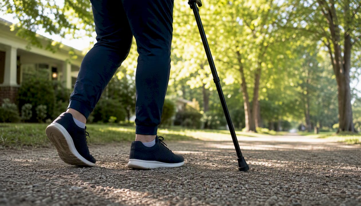
Activity modifications that exploit canal dynamics are practical and underutilized. Leaning slightly forward while walking, using a walking aid, choosing cycling over treadmill exercise, and taking brief seated rest breaks during longer walks can meaningfully extend functional walking distance while conservative care is underway.
Minimally invasive procedures and open surgery: matching the option to the patient
Surgery is considered when conservative care has failed over a reasonable trial period, typically three to six months, or when neurologic deficits are progressive or red flags are present. The choice of procedure depends on the severity and location of stenosis, the presence of instability, and the patient’s overall health and functional goals.
| Procedure | Best suited for | Expected relief / timeline | Key risks | Recovery |
|---|---|---|---|---|
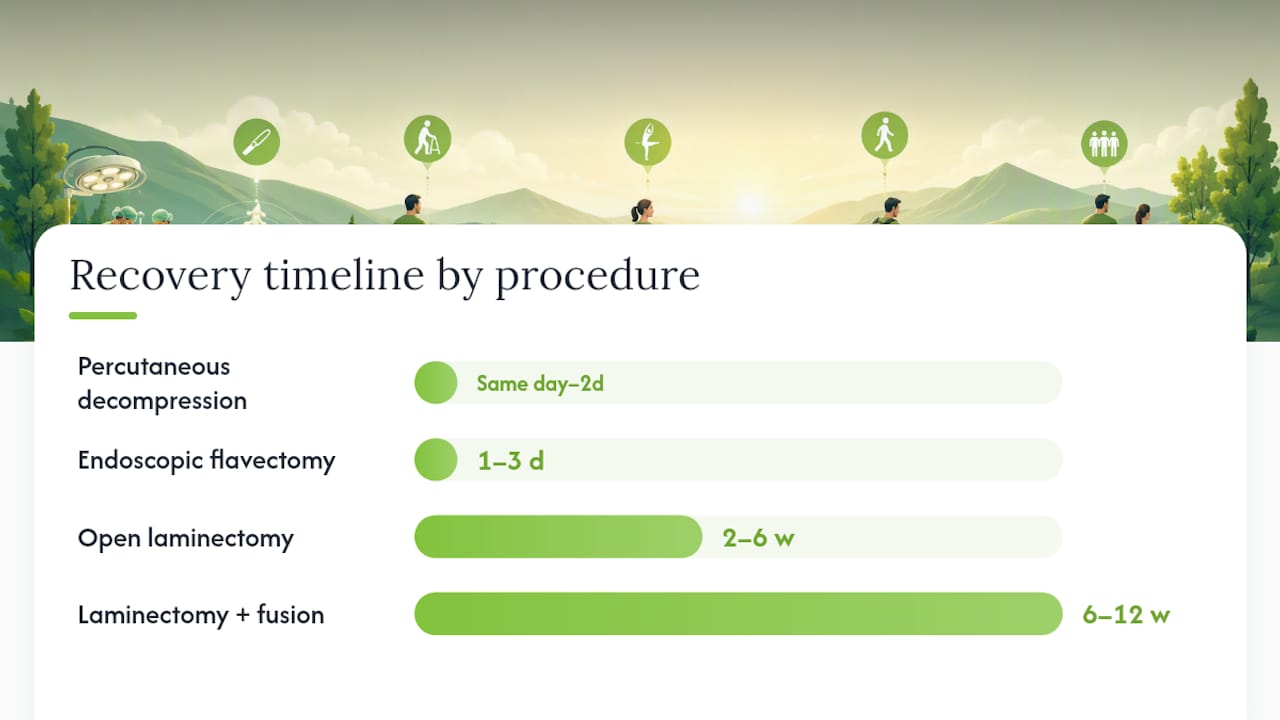
| Percutaneous lumbar decompression | Mild to moderate central LFH, no instability, outpatient candidate | Gradual improvement over weeks; variable durability | Incomplete decompression, procedural discomfort | Same day to 1–2 days |
| Endoscopic flavectomy / tubular microdecompression | Moderate LFH with or without lateral recess stenosis, no significant instability | Meaningful relief within days to weeks | Dural tear, nerve injury, recurrence | 1–3 days; return to light activity in 1–2 weeks |
| Open laminectomy (decompression) | Moderate to severe stenosis, multilevel disease, failed minimally invasive approach | Substantial relief in most patients; durable at 2–4 years | Infection, dural tear, instability, adjacent segment disease | 2–6 weeks; full recovery 3–6 months |
| Laminectomy with fusion | Stenosis with concurrent spondylolisthesis or instability | Similar decompression relief; fusion adds stability | Higher complication rate, longer OR time, pseudarthrosis | 6–12 weeks; full recovery 6 months |
Percutaneous lumbar decompression is an outpatient option for appropriately selected patients with hypertrophied ligamentum flavum and no significant instability. Minimally invasive decompression techniques, including endoscopic flavectomy and tubular microdecompression, target the ligament specifically with less collateral tissue trauma than open surgery, though candidate selection and surgeon experience are key determinants of success.
Open laminectomy remains the reference standard for moderate to severe stenosis and produces durable relief in most patients. Fusion is added when instability, spondylolisthesis, or significant facet destruction is present. Understanding post-laminectomy outcomes and potential complications is an important part of the preoperative discussion.
Outcomes data consistently show that patients with shorter symptom duration, preserved baseline function, and isolated LFH without multilevel disease tend to do better after decompression than those with longstanding deficits or significant comorbidities.
What to expect after treatment: outcomes and the image-mismatch problem
Many patients improve meaningfully with conservative care, particularly when symptoms are mild to moderate and neurologic deficits are absent. Physical therapy and injections typically produce benefit over weeks to a few months, though the structural thickening remains. For patients who proceed to minimally invasive decompression, functional recovery often begins within days to a few weeks. Open surgery carries a longer recovery arc: most patients return to light activity within two to six weeks, with full recovery taking three to six months depending on the extent of the procedure.
Predictors of better outcomes:
- Shorter duration of symptoms before treatment.
- Preserved baseline walking capacity and neurologic function.
- Isolated LFH at one or two levels without significant multilevel disease.
- Absence of significant comorbidities (diabetes, peripheral vascular disease) that impair nerve recovery.
Predictors of less favorable outcomes:
- Longstanding neurologic deficits (weakness, sensory loss) that may not fully reverse even after adequate decompression.
- Multilevel stenosis requiring extensive surgery.
- Concurrent spondylosis or significant facet arthropathy contributing to residual pain after decompression.
- Psychosocial factors and poor baseline functional status.
The symptom-image mismatch deserves emphasis. Clinical guidance consistently notes that many older adults have imaging changes that are asymptomatic, and that clinical decision-making should prioritize function and objective neurologic findings over raw MRI measurements. A patient who walks two blocks before stopping due to leg pain and weakness is a surgical candidate regardless of whether the ligament measures 4.5 mm or 6 mm. Conversely, a patient with a 5 mm ligament and no functional limitation does not need decompression.
When should you see a spine specialist?
Seek urgent evaluation, same day or through an emergency department, if you develop any of the cauda equina red flags described earlier: bilateral progressive leg weakness, saddle anesthesia, or new bowel or bladder dysfunction. These findings require immediate imaging and likely surgical consultation.
Outside of red flags, see a spine specialist if you experience any of the following:
- Leg pain, numbness, or weakness that limits your walking to less than one to two blocks.
- Symptoms that have not improved after six to eight weeks of consistent conservative care.
- Progressive neurologic findings on examination (worsening weakness, new reflex changes).
- Diagnostic uncertainty, where your symptoms do not clearly match your imaging findings.
What the specialist will do: Correlate your MRI findings with a detailed physical examination, order dynamic imaging if positional instability is suspected, review prior treatments, and discuss the trade-offs of continued conservative care versus procedural or surgical options. Bring a timeline of your symptoms, a list of treatments you have already tried and their results, and copies of any prior imaging studies or reports.
Pro Tip: If a surgeon recommends fusion at the first consultation without discussing a decompression-only option, asking specifically whether decompression alone is appropriate for your anatomy is reasonable. A second opinion before elective spinal fusion is standard practice and most spine specialists expect it.
How Nortexspineandjoint approaches LFH in clinical practice
At Nortexspineandjoint, the evaluation of a patient with suspected LFH follows a stepwise, evidence-informed pathway. The first priority is correlating the MRI findings with your actual functional limitations and neurologic examination, because the image alone does not determine the treatment plan.
Our clinical pathway:
- Step 1: Detailed history and physical examination to quantify functional walking distance, identify neurologic deficits, and assess red flags.
- Step 2: Review of existing imaging; order dynamic MRI or standing X-rays when instability or positional symptoms suggest they are needed.
- Step 3: Focused conservative care, including physical rehabilitation, anti-inflammatory management, and targeted epidural or facet injections when indicated.
- Step 4: For patients with inflammatory or fibrotic features who have not responded adequately to standard conservative care, regenerative options including PRP therapy for back pain and bone marrow biologics may be considered as adjuncts to reduce local inflammation and support tissue environment, with realistic expectations that these approaches address the inflammatory component rather than reverse established fibrosis.
- Step 5: When conservative and regenerative options are insufficient, we coordinate minimally invasive procedural options or surgical referral with a trusted spine surgeon, ensuring the patient understands the trade-offs at each stage.
Many patients come in after trying multiple treatments elsewhere without a clear explanation of why their symptoms behave the way they do. Taking the time to explain the canal dynamics, why flexion relieves symptoms, and what the realistic trajectory of each option looks like tends to improve both adherence and outcomes.
This information is general in nature and does not substitute for individualized medical evaluation. Treatment decisions should be made in consultation with a qualified clinician who has reviewed your specific imaging and examination findings.
A clinician’s perspective on treating LFH
What I find most important in managing ligamentum flavum hypertrophy is resisting the pull of the MRI number. Patients often arrive convinced that a thick ligament means they need surgery, or conversely, that a “borderline” measurement means nothing is wrong. Neither is reliably true.
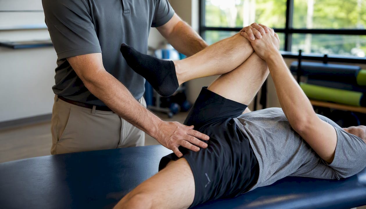
The patients who do best, regardless of the treatment path, are those who understand that this is a degenerative process with a spectrum of severity, and that the goal is restoring function rather than normalizing an image. Some patients with significant thickening do beautifully with a structured physical therapy program and one or two targeted injections. Others with similar imaging findings have progressive weakness that makes early surgical decompression the right call. Shared decision-making, with honest discussion of realistic timelines and the limits of each option, is what guides those choices well.
Nortexspineandjoint’s services for patients with LFH
If your MRI shows ligamentum flavum thickening and you are trying to determine the right next step, Nortexspineandjoint offers a structured evaluation process at our North Dallas clinic that goes beyond reviewing the image. We assess your functional walking capacity, neurologic status, and prior treatment history before recommending any intervention.
Relevant services include targeted epidural and facet injections, physical rehabilitation with flexion-based conditioning protocols, PRP therapy and biologic injections for patients with inflammatory or early fibrotic features, and coordination of minimally invasive or surgical options when the clinical picture warrants escalation. Our approach is evidence-informed and patient-first: we do not recommend procedures that the clinical picture does not support, and we set realistic expectations at every stage.
To request an evaluation, contact our clinic directly. Bring your most recent MRI report and images, a written timeline of your symptoms, and a list of treatments you have already tried. We will review everything before your appointment so the consultation focuses on your specific situation and next steps.
Sources
- Lumbar Spinal Stenosis – StatPearls – NCBI Bookshelf
- A novel rabbit model demonstrating mechanical stress and TGF‑β1 involvement in ligamentum flavum hypertrophy
- The Role of the Ligamentum Flavum Area as a Morphological Parameter of Lumbar Central Spinal Stenosis
- Radiopaedia
This article is general information, not a substitute for advice from a qualified doctor. Consult a qualified healthcare professional about your own circumstances before acting on anything here.
FAQ
Is ligamentum flavum hypertrophy serious?
It can be, depending on the degree of canal narrowing and your neurologic status. Many people have mild thickening without significant symptoms, while others develop progressive neurogenic claudication or radiculopathy that limits daily function and may eventually require surgical decompression.
What is the surgical treatment for ligamentum flavum hypertrophy?
The standard surgical approach is lumbar laminectomy with flavectomy, which removes the hypertrophied ligament and decompresses the spinal canal. Minimally invasive options including endoscopic flavectomy and tubular microdecompression are available for appropriately selected patients; fusion is added when instability or spondylolisthesis is also present.
Is ligamentum flavum hypertrophy a disability?
LFH itself is not automatically classified as a disability, but the functional limitations it causes, particularly severe neurogenic claudication that restricts walking to short distances, can qualify as a disabling condition under Social Security and other disability frameworks depending on severity and response to treatment.
Can physical therapy help ligamentum flavum hypertrophy?
Yes, physical therapy is a first-line treatment. Flexion-based conditioning, core strengthening, and gait training can meaningfully reduce symptoms and improve walking tolerance by optimizing canal dynamics, even though PT does not reverse the underlying fibrotic thickening of the ligament.
How is LFH different from a herniated disc?
A herniated disc compresses nerve roots from the front of the canal through disc material; LFH compresses from the posterior wall via the thickened ligament. Both can cause radicular symptoms, but LFH more typically produces bilateral neurogenic claudication with positional relief, while disc herniation more often causes unilateral radiculopathy that is worse with sitting or forward bending.